
Osteoarthritis: What happens in the hip joint?
Osteoarthritis of the hip: How to understand symptoms, causes and progression in X-ray images

Osteoarthritis as a widespread disease
Osteoarthritis is one of the most common degenerative joint diseases worldwide. The hip joint is particularly often affected, as it is subjected to high mechanical stress in everyday life. But what exactly happens in the hip joint during osteoarthritis? How does the disease develop, and what signs of osteoarthritis can be recognized in the different stages? This article examines the individual stages of development of hip osteoarthritis and discusses the respective changes seen in X-ray images.
Healthy cartilage and its protective function
Cartilage plays a crucial role in the function and longevity of the hip joint. It acts as a shock absorber and ensures smooth movement of the joint surfaces. Its smooth surface reduces friction and distributes the load evenly across the underlying bone.
Properties of healthy cartilage:
- High water content for elasticity and pressure distribution
- Collagen fibers for stability
- No blood vessels or nerves, therefore insensitive to pain
As soon as this cartilage is damaged, the degenerative process of osteoarthritis begins.
First phase of osteoarthritis: cartilage changes and reduction in cartilage thickness
In the early stages of osteoarthritis of the hip, severe symptoms usually do not yet occur. However, changes are already taking place in the joint:
- The cartilage loses water content and becomes less elastic.
- The first fine cracks appear in the cartilage layer.
- The cartilage thickness decreases, leading to uneven stress.
Early signs of osteoarthritis:
- Morning stiffness of the hip joint
- Slight limitation of movement
- Pain during exertion that subsides after periods of rest
Second phase of osteoarthritis: sclerosis of the bone and osteophyte formation
As the cartilage wears down, the underlying bone is subjected to increased stress. The body reacts to this with a process called sclerosis, a thickening of the bone to make it more resilient.
At the same time, the body tries to increase the surface area of the joint in order to better distribute the load. This leads to the formation of osteophytes (bone spurs) .
Symptoms of osteoarthritis at this stage:
- Pain during everyday movements
- Limitations in hip mobility
- Noticeable change in joint shape
X-ray signs of osteoarthritis:
- Joint space narrowing
- Increased density of the bone beneath the cartilage (subchondral sclerosis)
- First osteophytes on the joint edges
Third phase of osteoarthritis: Formation of subchondral cysts
subchondral cysts develop in the deeper bone layer . These are cavities filled with fluid or dead tissue. They arise from microfractures that the body can no longer properly repair.
Consequences of rock cysts:
- The bone loses stability
- The joint is becoming increasingly unstable
- The pain continues to increase
End stage of osteoarthritis: collapse of the joint surface and avascular necrosis of the femoral head
avascular necrosis of the femoral head develops , in which the bone is no longer adequately supplied with blood and dies.
Symptoms of end-stage osteoarthritis:
- Severe pain, even at rest
- Significant restriction of movement
- Changes in gait, even to the point of limping
X-ray signs of osteoarthritis:
- Severe deformation of the femoral head
- Almost complete loss of joint space
- Large bony cysts in the bone
Treatment options for coxarthrosis
Depending on the stage of osteoarthritis, there are different treatment approaches:
Conservative treatment of osteoarthritis of the hip:
- Physiotherapy to improve mobility
- Medications to reduce pain and inflammation
- Hyaluronic acid injections
- Weight reduction to relieve the joint
Surgical treatment of osteoarthritis of the hip:
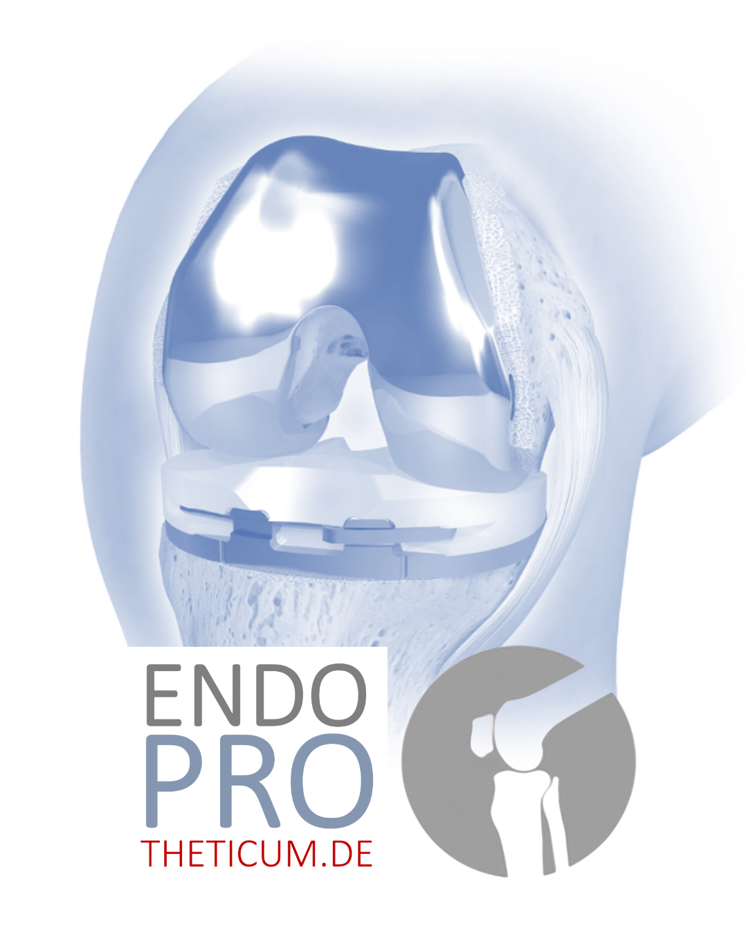
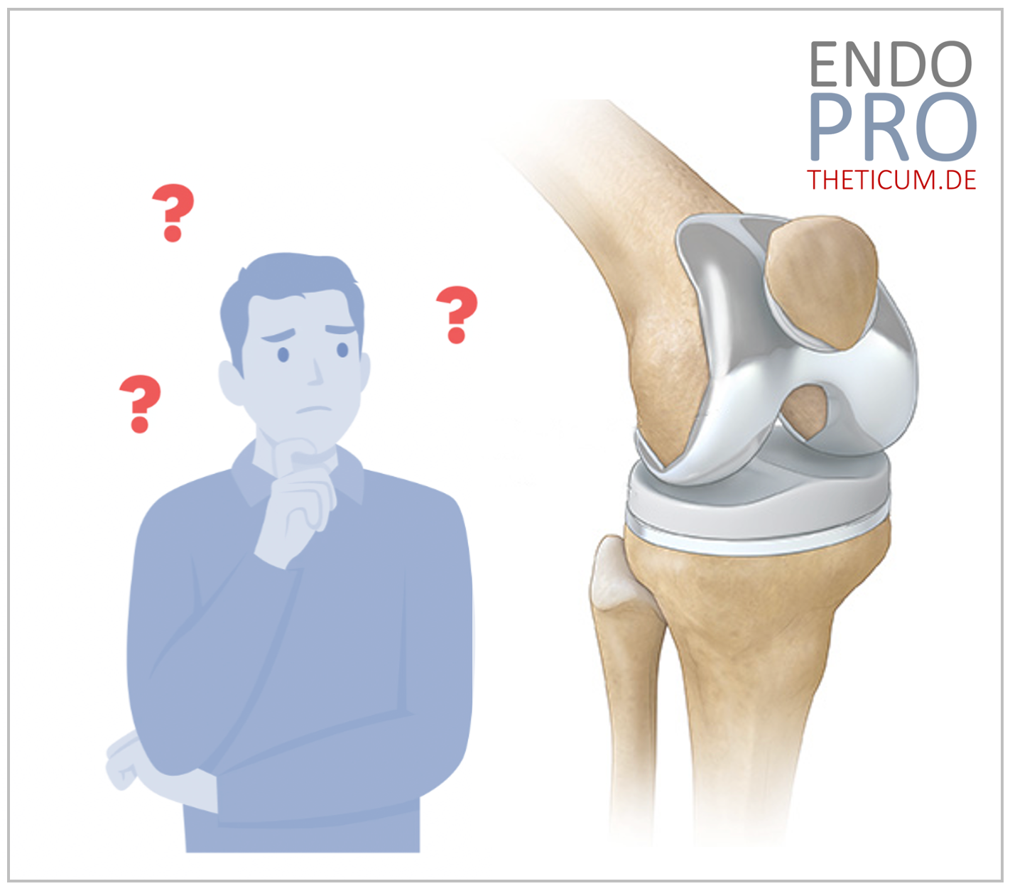
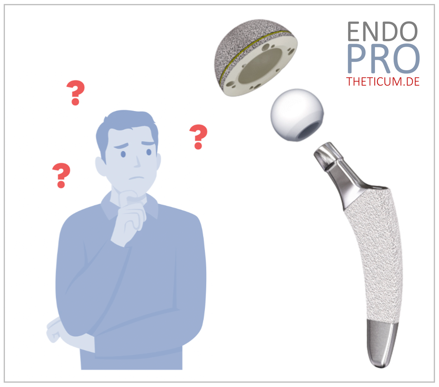
If conservative treatments are no longer sufficient, a total hip replacement (THR) be necessary. In this procedure, the damaged joint surfaces are replaced with a prosthesis that restores the original function of the joint.
Conclusion: Recognizing and treating hip osteoarthritis
Hip osteoarthritis is a gradual process that develops over years. The first signs of osteoarthritis are often nonspecific, which is why early diagnosis is crucial. Hip osteoarthritis is a progressive disease that, if left untreated, can lead to severe pain and significant limitations in movement. The typical signs of osteoarthritis, such as cartilage wear, bone sclerosis, osteophyte formation, and subchondral cysts, are clearly visible on X-rays and reflect the progression of the disease. While conservative measures such as physiotherapy, weight loss, and pain medication can be helpful in the early stages, in advanced stages, hip replacement surgery is often the only permanent solution.
Because the development of osteoarthritis varies from person to person and several factors must be considered, early consultation with a hip specialist is crucial. An experienced specialist can not only make a precise diagnosis but also develop a tailored treatment strategy – whether through joint-preserving measures or determining the optimal time for a hip replacement. Anyone suffering from hip pain should not wait too long but seek advice at a specialized center to find the best possible therapy for a pain-free future.
MAKE AN APPOINTMENT?
You are welcome to make an appointment either by phone or online .


ENDOPROTHETICUM - The whole world of endoprosthetics