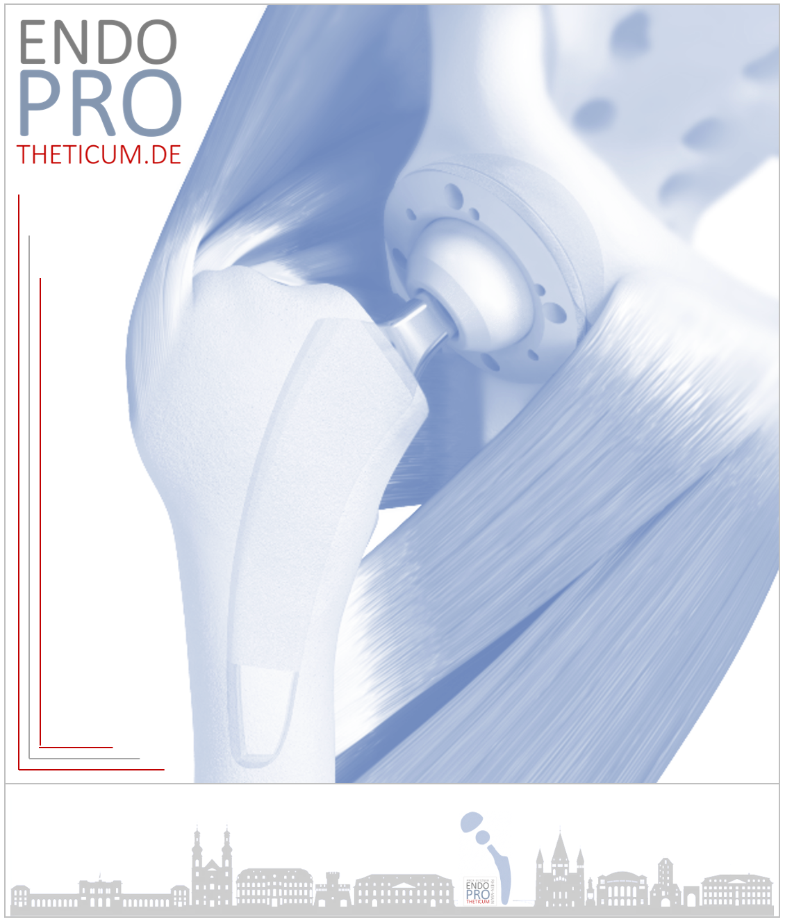
Skeletzestgraphy in diagnostics of the prosthesis loosening
Why a skeletal scintigraphy for diagnosing prosthesis loosening is only useful after approximately 1.5 years

Skeletal scintigraphy is an imaging technique based on the principle that radioactive markers (usually technetium-99m-labeled phosphates) are stored in areas of increased bone activity. This accumulation allows for the visualization of inflammation, infections, bone remodeling processes, or loosening of implants such as hip replacements (total hip arthroplasty) or knee replacements (total knee arthroplasty).
particularly helpful in diagnosing prosthesis loosening – however, there is an important time aspect:
1. Increased activity immediately after implantation
In the first 12 to 18 months after prosthesis implantation, a bone scan
always increased
uptake around the implant.
Why?
- After the prosthesis is inserted, the bone reacts to the trauma from the operation.
- Repair processes are taking place: bone remodeling and reaction to the new mechanical load.
- Additionally, micro-movements occur between the bone and the prosthesis until the final healing process is complete.
➡️ Result : Skeletal scintigraphy is almost always positive during this period – even if there is no loosening.
This means:
👉 Increased scintigraphy activity in the first 1 to 1.5 years is not specific for prosthesis loosening.
2. Risk of misinterpretation (false-positive findings)
Performing a scintigraphy scan during this period would pose a high risk for:
- False-positive findings : Normal healing or remodeling processes are mistakenly interpreted as loosening.
- Unnecessary interventions : Based on an incorrect diagnosis, unnecessary revision surgeries could be performed.
Example :
A patient experiences mild pain 10 months after total hip replacement surgery. A scintigraphy scan shows increased uptake. Without considering the healing process, loosening of the implant could be mistakenly assumed – however, this finding is completely normal for this phase.
3. Time point after 1.5 years – why this period?
After about 15–18 months :
- Renovation activity has largely returned to normal.
- The bone-implant interaction stabilizes.
- If an increased accumulation persists, this is indeed indicative of pathological processes (e.g., loosening, infection).
➡️ From this point on, skeletal scintigraphy has a significantly higher specificity and informative value for:
- Relaxations (aseptic or septic)
- Implant loosening due to infection or mechanical instability
- Bone damage or osteolysis
In short:
👉 Only after about 1.5 years can a scintigraphy reliably distinguish a true loosening from normal remodeling processes.
4. Current scientific consensus
Studies and guidelines (e.g., from the German Society for Orthopedics and Trauma Surgery, DGOU) therefore recommend:
- No skeletal scintigraphy for loosening diagnosis within the first 12–18 months after primary implantation of a hip replacement or knee replacement.
- Instead, during this period: Clinical examination, conventional X-ray diagnostics, possibly puncture in case of suspected infection.
5. Alternative diagnostics in the first 18 months
If loosening of a hip or knee prosthesis is suspected early on (e.g., in cases of persistent pain, fever, or functional limitations), the following should be used preferentially:
- Conventional X-ray (direct signs such as prosthesis loosening or osteolysis)
- CT (fine diagnostics, implant loosening test)
- Puncture (to differentiate between septic and aseptic loosening)
- Laboratory values (CRP, white blood cell count)
Only if these procedures remain unclear and the symptoms persist, should a scintigraphy be considered after approximately 18 months.
Conclusion: Skeletal scintigraphy in cases of prosthesis loosening is usually only useful after 1.5 years!
Skeletal
scintigraphy loosening of a hip or knee prosthesis suspected
. However, the timing must be considered:
In the first 12 to 18 months after implantation, the findings are often distorted by normal healing and remodeling processes, making interpretation difficult and prone to error.
Only about 1.5 years after surgery does bone scintigraphy provide meaningful evidence of actual loosening or a possible infection.
Therefore, it is crucial to use classic diagnostic methods such as clinical examination, X-ray diagnostics, and, if necessary, joint aspiration early on.
A targeted scintigraphy, used appropriately in terms of timing, can then make a crucial contribution to reliably diagnosing prosthesis loosening and setting the course for successful therapy.
Note:
🔵 1.5 years ago → Scintigraphy hardly usable due to normal healing.
🟢 After 1.5 years → Meaningful tool if loosening is suspected.
MAKE AN APPOINTMENT?
You are welcome to make an appointment either by phone or online .

ENDOPROTHETICUM - The whole world of endoprosthetics