Knee pain: Always think about your hips too!
Pain is transmitted from the hip to the knee via the iliotibial band

Knee pain is a common condition that affects people of all ages. It is often assumed that the cause of the pain lies directly in the knee joint. But medical examinations repeatedly show that the hip plays a central role in the development of knee pain. Radiating pain between the hip and knee joints is not uncommon and can make diagnosis significantly more difficult.
In this article, we take a detailed look at the connection between knee pain, hip disease, and the mechanisms of pain referral. We shed light on medical backgrounds, treatment options and preventative measures.
The complex connection between the knee and hip
Anatomical basics
The knee and hip joints are part of the lower extremity and work closely together to ensure the mobility and stability of the human body. Problems in the hip area can lead to incorrect biomechanical stress, which has a negative effect on the knee.
- Nerve pathways and pain radiation: The nerves that supply the hip overlap with those of the knee. Pain from the hip is therefore often “projected” and felt in the knee.
- Biomechanics: Misalignments or diseases of the hip change the gait and can increase the load on the knee joint, which leads to overloading or wear and tear.
Radiation of pain: How hip problems become noticeable
- Hip joint arthrosis: One of the most common causes of pain radiating to the knee. Stress-related pain is typical and is first felt in the knee.
- Hip impingement: Limited hip mobility can strain the iliotibial band muscle and secondarily lead to knee pain.
- Damage to the lumbar spine: Pain perception in the spine, hips and knees often overlaps due to the same nerve pathways.
The role of the iliotibial band in the transmission of pain from the hip to the knee
The iliotibial band , a broad tendon plate on the outside of the thigh, plays a central role in the power transmission and stability of hip and knee joint movements. Due to its connection to the hip muscles and the knee, it can be both the cause and the path for the transmission of pain.
Anatomical connections
The iliotibial tract arises as a thickening of the fascia lata on the iliac crest. It is associated with several important muscle groups:
- Tensor fasciae latae (TFL) muscle: A hip flexor that tightens the tract and provides stability during gait movement.
- Gluteus maximus muscle: Also supports the tension of the iliotibial band.
At the knee, the tract attaches to Gerdy's tubercle (lateral tibial condyle) and stabilizes the joint in extension and flexion.
Mechanisms of pain transmission
Pain radiating from the hip to the knee can be mediated via the iliotibial band through the following mechanisms:
- Increased tension in the iliotibial band:
- Causes such as hip misalignments, osteoarthritis or muscular imbalances can overstrain the tract.
- Chronic tension transfers friction or tension to its distal attachments to the knee joint, resulting in pain in the lateral knee area.
- Inflammation of the iliotibial band (IT band syndrome):
- Repetitive movements, such as running or climbing stairs, cause friction between the tract and the lateral femur (via the lateral epicondyle).
- This is often misinterpreted as an isolated knee problem, but often has a primary cause in hip mechanics.
- Myofascial pain transmission:
- Trigger points in the muscles, especially the tensor fasciae latae or gluteus maximus muscles, can radiate along the path of the iliotibial band to the knee.
Causes and triggers in the hip area
Common triggers of overuse of the iliotibial band that cause secondary knee pain include:
- Hip dysplasia: Causes altered loading patterns in the lateral thigh structures.
- Hip impingement: Restrictions in hip mobility lead to compensatory overstressing of the tract.
- Pelvic misalignments: Unbalanced tension in the tract due to asymmetrical pelvic position.
Common diseases related to the hip and knee
Osteoarthritis: When the hip affects the knee
Hip osteoarthritis (coxarthrosis) can lead to changes in gait and increased stress on the knee. Increased limping or a protective posture puts excessive strain on the knee joint and accelerates the wear and tear process.
Hip bursitis
Inflamed bursa (“bursitis”) on the hip often causes local pain that can radiate to the knee. The similar symptoms can lead to misdiagnosis.
Herniated discs
A herniated disc in the lumbar spine can put pressure on the sciatic nerve and cause pain that radiates to both the hip and knee.
Knee pain typical of hip dysplasia
Hip dysplasia is a congenital or early-childhood-acquired malformation of the hip joint in which the acetabulum inadequately covers the femoral head. This improper fit leads to instability of the hip joint, which can cause a variety of long-term mechanical and nerve problems, including knee pain.
Mechanisms that explain knee pain in hip dysplasia:
- Biomechanical effects:
- The unstable hip disrupts the transfer of force while walking. The load is often shifted to the knee joint, which can lead to incorrect loading and pain in the knee.
- Compensatory changes in the leg axis often occur, which lead to unequal loading of the medial or lateral knee joint.
- Radiation of pain:
- The hip joint shares a common sensory supply with the knee, particularly via the femoral nerve and obturator nerve. This means that pain from the hip area can be perceived as knee pain.
- Gait pattern abnormalities:
- Patients with hip dysplasia tend to adapt their gait through posture or increased pelvic movements. This increases the overload on the knee region.
Diagnostic strategies: Finding the origin of the pain
Clinical examination
- Gait analysis: Abnormalities such as limping or shortened steps provide information about the strain on the hips and knees.
- Palpation: Pain when pressure is applied to specific areas helps narrow focus.
Imaging procedures
- X-ray images: Ideal for detecting osteoarthritis or fractures.
- MRI: Provides detailed images of the soft tissues and helps diagnose bursitis or muscle injuries.
- Ultrasound: Particularly useful for visualizing swelling or effusions.
Therapeutic approaches: Multidisciplinary solutions
Conservative treatment
- Physiotherapy: The aim is to strengthen and stabilize the muscles in order to eliminate biomechanical imbalances.
- Pain management: NSAIDs (non-steroidal anti-inflammatory drugs) help reduce acute inflammation.
- Orthopedic aids: Individually adjusted shoe inserts or bandages relieve the affected joint.
Surgical interventions
In some cases, especially with advanced osteoarthritis or serious injuries, operations are required:
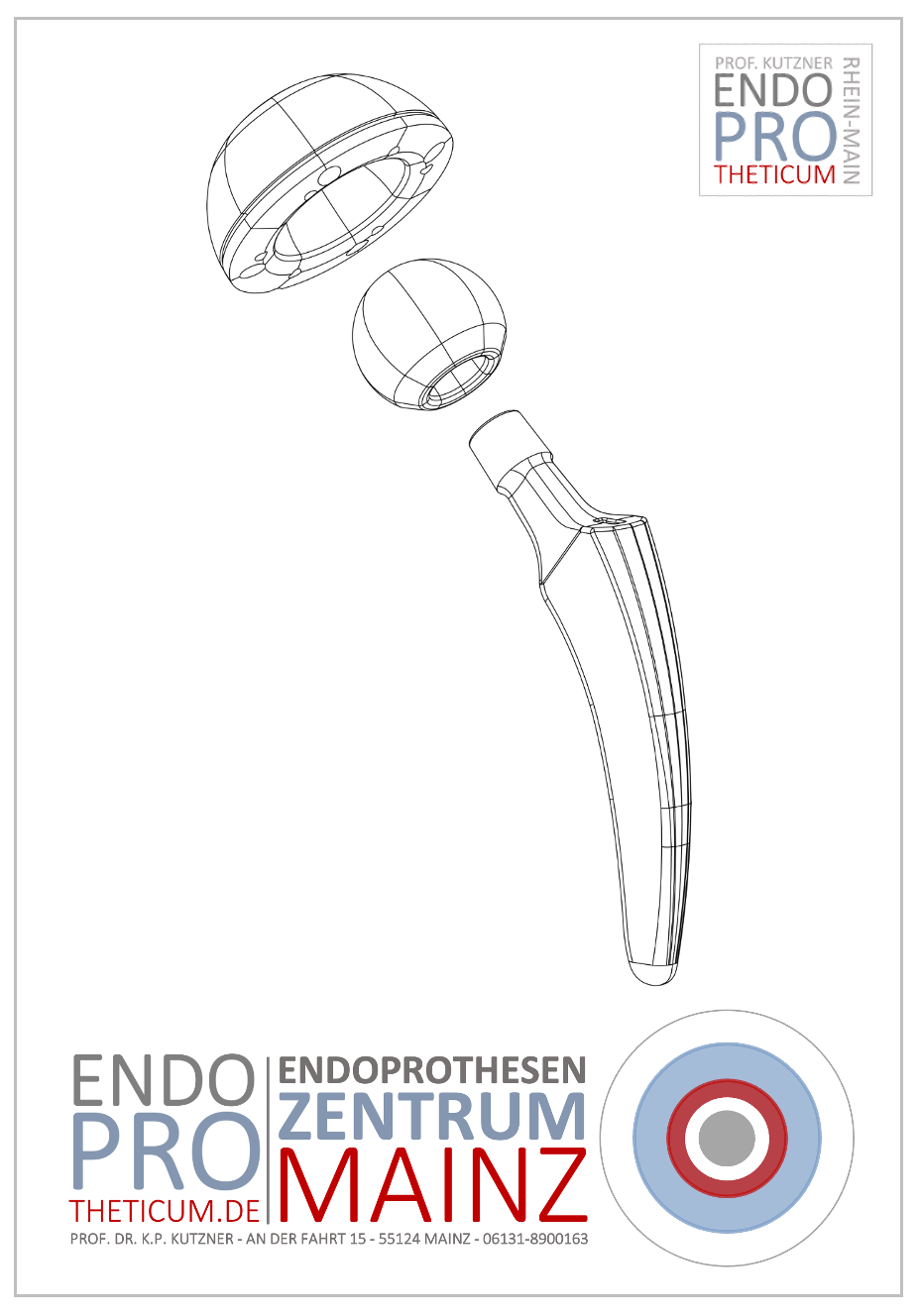
- Hip prosthesis: Replacement of the damaged hip joint reduces secondary knee pain.
- Arthroscopy: Minimally invasive procedures can correct changes in the hip joint.
Prevention: How to prevent knee pain with healthy hips
Regular exercise
A balanced training program that combines strength, flexibility and endurance protects both joints.
Ergonomics in everyday life
- Sitting correctly: Avoiding sitting in a hunched position for long periods of time.
- Hip-friendly strain: Regularly change sitting and standing positions.
Nutrition
A diet rich in calcium and vitamin D strengthens bones and joints.
Conclusion
Knee pain should always be viewed holistically, as it often originates from the hip or other anatomical structures. An accurate diagnosis and collaboration between various specialists are crucial for successful treatment.
By improving awareness of these connections, many patients can receive appropriate therapy earlier and avoid long-term complications.
MAKE AN APPOINTMENT?
You are welcome to make an appointment either by phone or online .


